
Learning Objectives
- Define the chromosome theory of inheritance as “genes are located on chromosomes” and know that chromosomes can be solo or paired with homologs that contain the same genes but possibly different gene variants, called alleles.
- Know that DNA is made up of four bases—A, G, T, and C—that spell out instructions to make proteins and recognize three types of mutations in DNA: insertion, deletion, and point mutation.
- Diagram and explain that for genes to be expressed they are transcribed from DNA to RNA, which is then translated from RNA into amino acid chains of protein (the central dogma of molecular biology).
- Examine a karyotype to predict information about the individual, including sex and presence of chromosomal conditions of Klinefelter, Turner, and Down syndrome. Be able to differentiate the three types of Down syndrome.
What we’ve learned already:
DNA is the universal information storage for all living organisms, and it is faithfully replicated before cell division to be passed from parent cell to the daughter cells. Accurate replication makes daughter cells identical to the parent cell.
Bacteria and archaea have single chromosomes that are ring-shaped, while eukarya have linear chromosomes. Eukaryotes often have multiple linear chromosomes. We’ve also established that some organisms are haploid, having one copy of each chromosome, while others are diploid and have two copies of each chromosome.
Chromosomes
A chromosome is one long chain of the genetic material DNA and can have hundreds to thousands of genes located along its length. To make proteins, genes spell out instructions using an alphabet of only four letters—A, C, T, and G—called bases. Most genes require at least a few hundred letters (bases) to encode a single protein, and eukaryotic chromosomes are quite long, several meters, but most cells are so much smaller than that. A meters long chromosome can fit inside a cell because it’s wound up tightly in in an organized way around special proteins (called histones in the diagram).
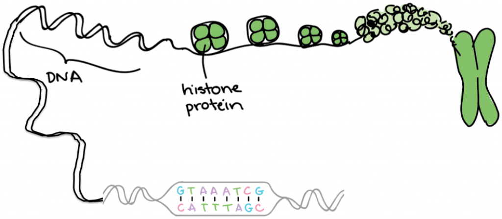
Image credit: Modified from Khan Academy.
As a result of DNA replication before cell division, replicated chromosomes consist of a pair of sister chromatids. Each chromatid contains a linear DNA molecule that is “identical” to the sister chromatid joined to it. We’ve put the word identical in quotes to emphasize an important point: DNA replication is pretty accurate, but not wholly accurate. In humans, errors in DNA replication occur only 1 in 100,000,000 bases (or 10^8). These errors are called mutations. A mutation is a change in the DNA from the original to the replicated copy.
While mutations are rare, they can be very important for the organism that inherits them. Replication can generate one of three types of mutations in DNA: insertion, deletion, and point mutation. An insertion mutation adds letters into the DNA code, similar to this typo: mutatation. A deletion mutation removes letters, like this: mutaton. A point mutation changes a letter, perhaps by turning mutation into tutation or mutasion. Sometimes these mutations break the gene (what’s a tutation, for instance?) while other times the change doesn’t really impact the organism (mutasion sounds almost the same as mutation). The point here is that some mutations are more harmful than others when the gene is eventually expressed as a protein.
DNA is transcribed into RNA, which is translated to form proteins
Francis Crick coined the phase “the Central Dogma” to describe the flow of information from nucleic acid to protein. Information encoded in DNA is transcribed to a similar alphabet called RNA (still four letters only, but not quite the same four letters as DNA). RNA is translated to a linear sequence of amino acids in protein. This video gives a concise overview of the central dogma of molecular biology:
We will go into more detail about gene expression later in the course.
Your chromosomes provide the instructions for how your body will develop before you’re born, and how it will function as you grow. Typically, humans are born with 23 pairs of chromosomes (46 chromosomes in total). Human chromosomes 1 through 22 are called autosomes, and the final or 23rd chromosome is a pair of sex chromosomes, so-called because they determine the biological sex of the human.

Recall that half of your chromosomes come from your mother and half come from your father. Through a process called meiosis each of your parents creates a sex cell (either a sperm or an egg) that contains half of their chromosomes. When the sperm cell and the egg cell combine, they create a zygote that contains 46 chromosomes.
Extra sex chromosomes and X-chromosome inactivation
Having extra or missing chromosomes in most cases is lethal to humans (causing an embryo to die early in development). For example, you’ve never met someone with an extra copy of chromosome 9 or 10; that seems to be a genotype that cannot exist. However, some combinations of extra chromosomes can not only exist but also thrive. For instance, Down Syndrome is caused by an extra copy of chromosome 21 (more on that below). The most common example of an extra copy of a chromosome is in XX females. Females have two X chromosomes (XX), while males have just one X (XY). Why doesn’t it cause problems for males to have just one copy of the X chromosome, while females have two?
The answer is so weird! In humans and many animals, the level of gene activity produced by a single X chromosome is considered the normal “dosage.” Males have this dosage automatically because they only have one X chromosome. Females have the gene activity of a single X for a different reason: even though they have XX in every cell, their cells switch off one of their two X chromosomes in a process called X-inactivation.
Females inherit two X chromosomes, one from each parent. Which one will be inactivated? In humans, and most mammals, X-inactivation is a random process that happens independently in every cell during embryonic development. One cell might switch off the paternal X, while the cell next to it might switch off the maternal X instead. All the cells descended from each of these original cells will maintain the same pattern of X-inactivation. So, humans are a mosaic of X-inactivation. But if you were a kangaroo, or any marsupial mammal, your paternal X chromosome would always undergo X-inactivation.
The inactivated X chromosome compacts or crumples into a small, dense mass called a Barr body. As we read above, having an extra or missing copy of a chromosome is unusual for most chromosomes. However, because of X inactivation, we see chromosomal conditions that involve extra X chromosomes more frequently. Although the purpose of the X-inactivation system is to switch off the second X of an XX female, it can also do a pretty good job of switching off more X chromosomes if they are present.
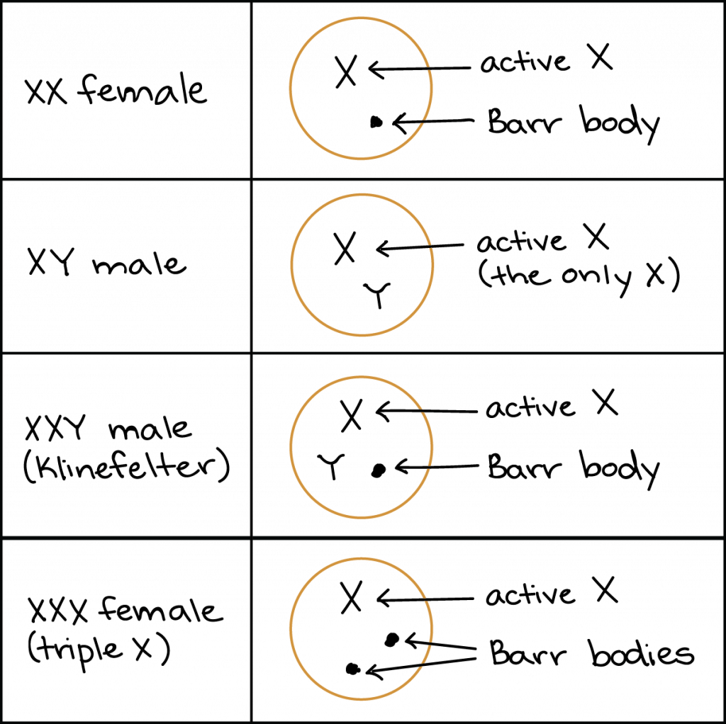
Image credit: Khan Academy.
Examples of X chromosome conditions include:
Triple X syndrome, in which a woman has an XXX genotype, which occurs in about 1 out of every 1000 female newborns. Women with an XXX genotype have female sex characteristics and are fertile (able to have children). In some cases, triple X syndrome may be associated with learning difficulties, late development of motor skills in infants, and problems with muscle tone.
Klinefelter syndrome, in which males have an extra X chromosome, leading to a genotype of XXY. (In rarer cases, Klinefelter syndrome can involve several extra Xs, leading to an XXXY or XXXXY genotype.) Affected men may be infertile or develop less dense body and facial hair than other men. Klinefelter syndrome is thought to affect 1 out of every 500-1000 male newborns.
Turner syndrome occurs when a woman lacks part or all of one of her X chromosomes, leaving her with just one functional X. Females with this condition often have short stature and may exhibit traits like infertility and learning difficulties. Turner syndrome is thought to occur in about 1 out of every 2500 female births. It has relatively mild effects because humans normally have only one X active in the cells of their body anyway.
Extra autosomes arise through uneven chromosome separation
Now let’s consider an autosomal condition with three copies of a chromosome: trisomy 21 or Down syndrome. Down syndrome is a chromosomal condition in which atypical cell division causes an extra portion of chromosome 21 to be present in some or all of a person’s cells.
What happens with Down syndrome?
During both mitosis and meiosis, there is a phase where each chromosome pair in a cell is separated, so that each new cell can get a copy of every chromosome. However, sometimes during this process, a pair of chromosomes doesn’t separate evenly, which results in one of the new cells having an extra section of chromosome. With Down syndrome, various types of uneven chromosome separation result in a person having an extra copy (or partial copy) of chromosome 21.
There are three main types of Down syndrome: trisomy 21, mosaicism, and translocation.
Trisomy 21
Trisomy 21 is the most common form of Down syndrome, accounting for about 95% of cases. This type of Down syndrome is caused by uneven separation of chromosome 21 during the creation of sex cells (this can happen in either the sperm or the egg cell), which leads to a fertilized egg with three copies of chromosome 21 instead of two. When the fertilized egg is developed, it passes along the extra copy of chromosome 21 to every cell in the body.

Diagram of genetic transmission of trisomy 21. Image credit: Khan Academy.
Mosaicism
The mosaic form of Down syndrome is much less common, accounting for about 1% of cases. In this form, the uneven separation of chromosome 21 happens shortly after an egg has been fertilized. The timing of when the chromosomes separate unevenly is important, because it leads to a person having some cells with the typical 46 chromosomes, and some cells with 47 (these cells have an extra copy of chromosome 21). Because only some cells have the extra chromosome, mosaic Down syndrome may have less impact than trisomy 21.
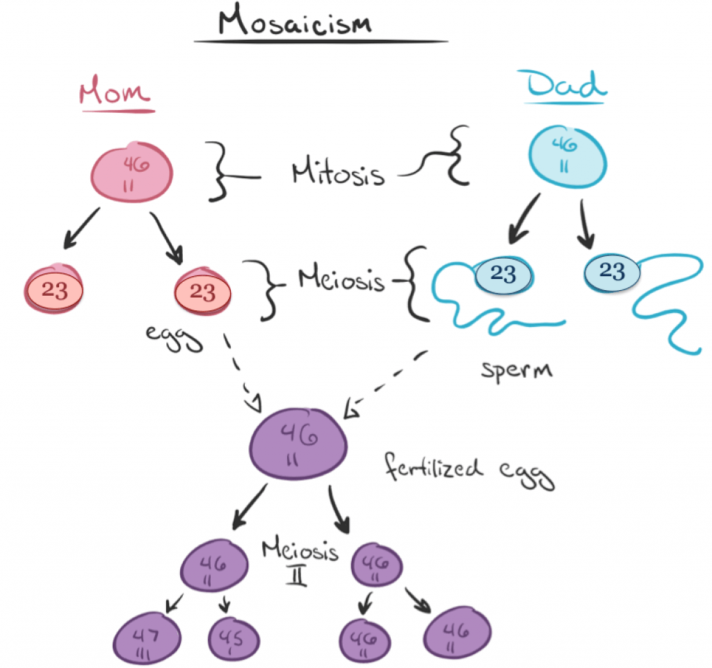
Mosaicism for Down syndrome. Image modified from Khan Academy.
Translocation
In the remaining 4% of cases of Down syndrome, the extra genetic material is passed on to new cells in a slightly different way. Rather than failing to separate, a portion of chromosome 21 breaks off during the replication process, and then attaches to another chromosome. So rather than getting a full extra chromosome, translocation results in cells with the typical 46 chromosomes, plus a little extra piece of chromosome 21. The genes contained in the extra portion of chromosome 21 can cause many of the traits of Down syndrome.